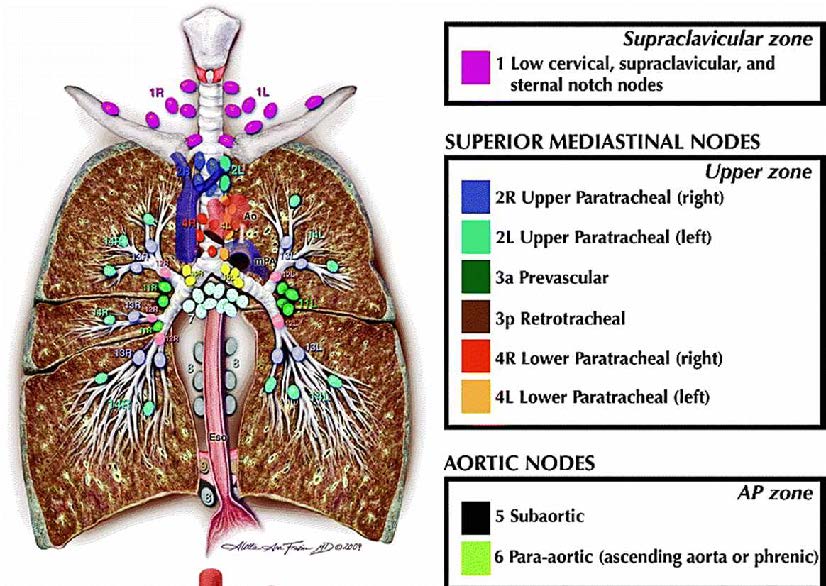
Supraclavicular nodes
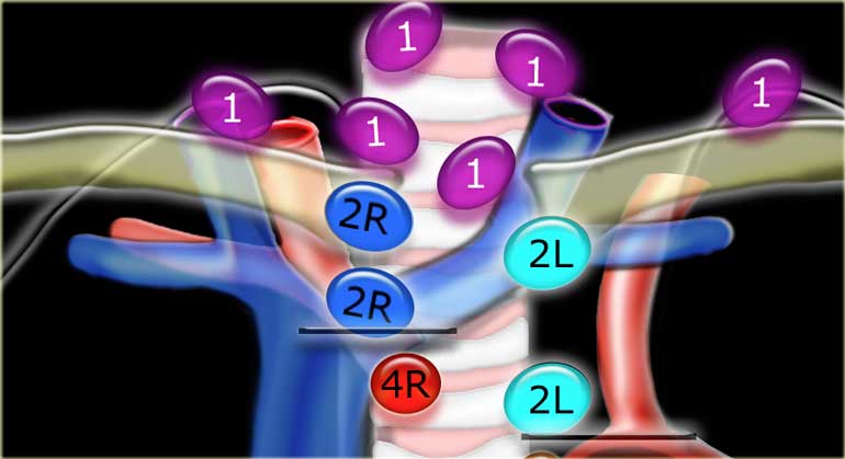
1.Low cervical, supraclavicular and sternal notch nodes
From the lower margin of the cricoid to the clavicles and the upper border of the manubrium.
The midline of the trachea serves as border between 1R and 1L.
Superior Mediastinal Nodes 2-4
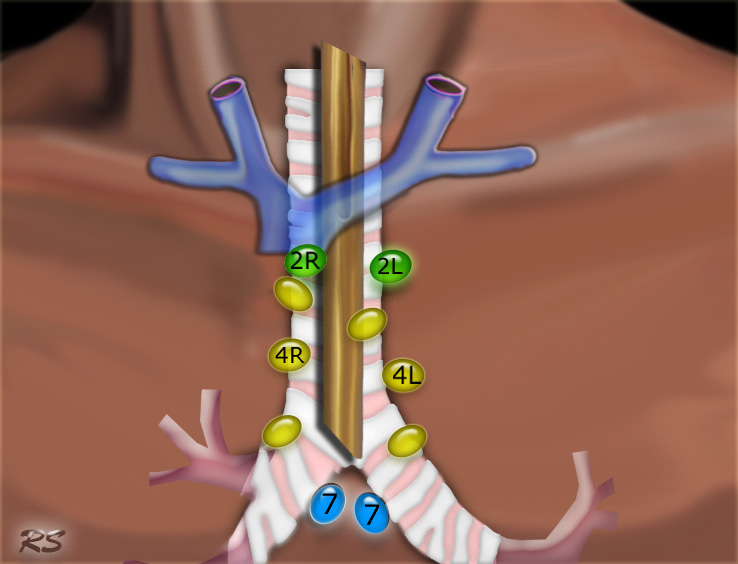
2R.Upper Paratracheal
2R nodes extend to the left lateral border of the trachea.
From upper border of manubrium to the intersection of caudal margin of innominate (left brachiocephalic) vein with the trachea.
2L.Upper Paratracheal
From the upper border of manubrium to the superior border of aortic arch.
2L nodes are located to the left of the left lateral border of the trachea.
3A.Pre-vascular
These nodes are not adjacent to the trachea like the nodes in station 2, but they are anterior to the vessels.
3P.Pre-vertebral
Nodes not adjacent to the trachea like the nodes in station 2, but behind the esophagus, which is prevertebral.
4R.Lower Paratracheal
From the intersection of the caudal margin of innominate (left brachiocephalic) vein with the trachea to the lower border of the azygos vein.
4R nodes extend from the right to the left lateral border of the trachea.
4L.Lower Paratracheal
From the upper margin of the aortic arch to the upper rim of the left main pulmonary artery.
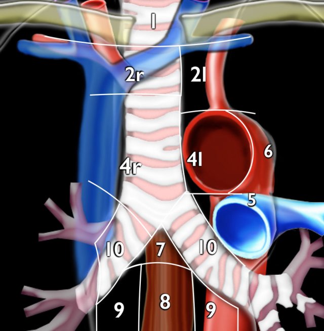
Aortic Nodes 5-6
- Subaortic
These nodes are located in the AP window lateral to the ligamentum arteriosum.
These nodes are not located between the aorta and the pulmonary trunk but lateral to these vessels. - Para-aortic
These are ascending aorta or phrenic nodes lying anterior and lateral to the ascending aorta and the aortic arch.
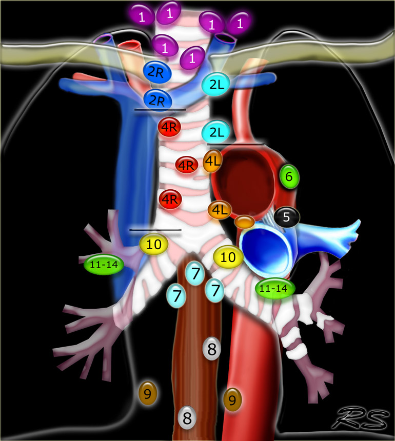
Inferior Mediastinal Nodes 7-9
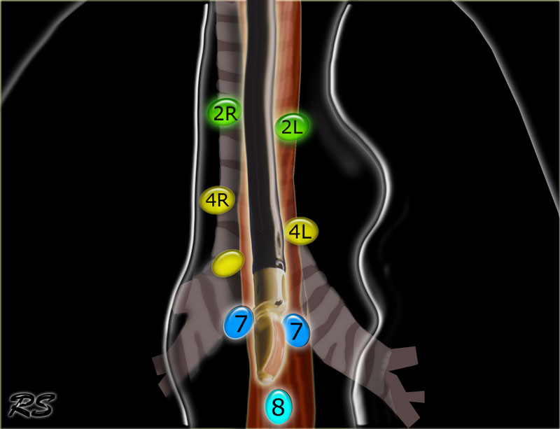
7.Subcarinal
- Paraesophageal
Nodes below carina. - Pulmonary Ligament
Nodes lying within the pulmonary ligaments.
Hilar, Lobar and (sub)segmental Nodes 10-14
These are all N1-nodes.
- Hilar nodes
These include nodes adjacent to the main stem bronchus and hilar vessels.
On the right they extend from the lower rim of the azygos vein to the interlobar region.
On the left from the upper rim of the pulmonary artery to the interlobar region.
- Supraclavicular zone nodes
These include low cervical, supraclavicular and sternal notch nodes.
Upper border: lower margin of cricoid.
Lower border: clavicles and upper border of manubrium.
The midline of the trachea serves as border between 1R and 1L.
2R. Right Upper Paratracheal
2R nodes extend to the left lateral border of the trachea.
Upper border: upper border of manubrium.
Lower border: intersection of caudal margin of innominate (left brachiocephalic) vein with the trachea.
2L. Left Upper Paratracheal
Upper border: upper border of manubrium.
Lower border: superior border of aortic arch.
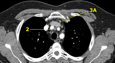
On the left a station 2 node in front of the trachea, i.e. a 2R-node.
There is also a small prevascular node, i.e. a station 3A node.
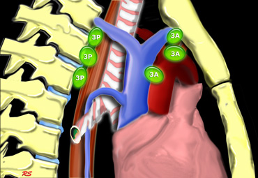
Prevascular and Prevertabral nodes
Station 3 nodes are not adjacent to the trachea like station 2 nodes.
They are either:
3A anterior to the vessels or
3B behind the esophagus, which lies prevertebrally.
Station 3 nodes are not accessible with mediastinoscopy.
3P nodes can be accessible with endoscopic ultrasound (EUS).

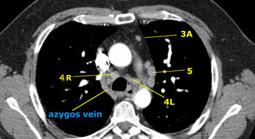
Right Lower Paratracheal
Upper border: intersection of caudal margin of innominate (left brachiocephalic) vein with the trachea.
Lower border:lower border of azygos vein.
4R nodes extend to the left lateral border of the trachea.
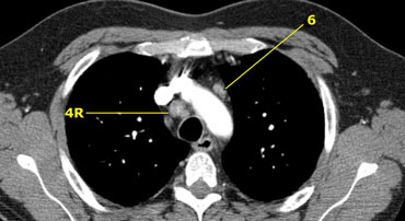
On the left we see 4R paratracheal nodes.
In addition there is an aortic node lateral to the aortic arch, i.e. station 6 node.

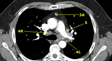
4L. Left Lower Paratracheal
4L nodes are lower paratracheal nodes that are located to the left of the left tracheal border, between a horizontal line drawn tangentially to the upper margin of the aortic arch and a line drawn tangentially to the upper margin of the left pulmonary artery.
These include paratracheal nodes that are located medially to the ligamentum arteriosum.
Station 5 (AP-window) nodes are located laterally to the ligamentum arteriosum.

- Subaortic nodes
Subaortic or aorto-pulmonary window nodes are lateral to the ligamentum arteriosum or the aorta or left pulmonary artery and proximal to the first branch of the left pulmonary artery and lie within the mediastinal pleural envelope. - Para-aortic nodes
Para-aortic (ascending aorta or phrenic) nodes are located anteriorly and laterally to the ascending aorta and the aortic arch from the upper margin to the lower margin of the aortic arch.


7.Subcarinal nodes
These nodes are located caudally to the carina of the trachea, but are not associated with the lower lobe bronchi or arteries within the lung.
On the right they extend caudally to the lower border of the bronchus intermedius.
On the left they extend caudally to the upper border of the lower lobe bronchus.
On the left a station 7 subcarinal node to the right of the esophagus.
8 Paraesophageal nodes
These nodes are below the carinal nodes and extend caudally to the diafragm.
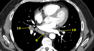
On the left an image below the carina.
To the right of the esophagus a station 8 node.
- Pulmonary ligament nodes
Pulmonary ligament nodes are lying within the pulmonary ligament, including those in the posterior wall and lower part of the inferior pulmonary vein.
The pulmonary ligament is the inferior extension of the mediastinal pleural reflections that surround the hila.
10 Hilar nodes
Hilar nodes are proximal lobar nodes, distal to the mediastinal pleural reflection and nodes adjacent to the intermediate bronchus on the right.
Nodes in station 10 - 14 are all N1-nodes, since they are not located in the mediastinum.

原文链接:http://www.xxwk.net/archives/1109